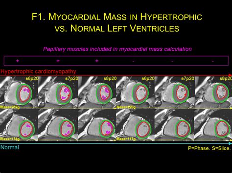
lv mass normal range | how to calculate Lv mass lv mass normal range This LV mass index calculator helps diagnose the type of cardiac hypertrophy based on LVMI and relative wall thickness. Omega kicked off the year 2021 with a bang by elevating the legendary Moonwatch to Master Chronometer. Although this watch is equipped with the latest .
0 · what is normal myocardial mass
1 · what is Lv mass 2d
2 · myocardial mass normal range
3 · left ventricular mass size 1
4 · left ventricular mass index chart
5 · how to calculate Lv mass
6 · Lv wall mass calculator
7 · Lv mass index chart
$9.99
versus versace arrondissement - chronograph watch
Visual assessment of regional wall motion (left ventricle) Recommended by American Society for Echocardiography (J Am Soc Echocardiogr 18:1440-1463, 2005). Left ventricular mass and geometry. Left ventricular dimension and .This LV mass index calculator helps diagnose the type of cardiac hypertrophy based on LVMI and relative wall thickness.
Normal values of left ventricular mass (LV M) and cardiac chamber sizes are prerequisites for the diagnosis of individuals with heart disease. LV M and cardiac chamber . Normal 2D measurements: LV minor axis ≤ 2.8 cm/m 2, LV end-diastolic volume ≤ 82 ml/m 2, maximal LA antero-posterior diameter ≤ 2.8 cm/m 2, maximal LA volume ≤ 36 ml/m 2 (2;33;35). ∗∗ In the absence of other .Normal Ranges for LV Size and Function Normal values for LV chamber dimensions (linear), volumes and ejection fraction vary by gender. A normal ejection fraction is 53-73% (52-72% for . Normal LV mass and LV mass index values established using linear, 2D, and 3D techniques stratified by sex are shown in Table 2. These numbers were derived from the entire .
versace women's round sunglasses
Left ventricular mass (LVM) is a well-established measure that can independently predict adverse cardiovascular events and premature death. 1-3 Population-based studies . For LV parameters counting papillary muscles and trabeculations in the LV mass, pooled normative reference ranges in men and women, respectively, were as follows: LV .We need to start reporting on LV Mass on all patients with hypertension. The normal reference range for LVM is gender specific and indexed to BSA. Concentric LVH = increased LVM; Eccentric LVH = increased LVM; .Visual assessment of regional wall motion (left ventricle) Recommended by American Society for Echocardiography (J Am Soc Echocardiogr 18:1440-1463, 2005). Left ventricular mass and geometry. Left ventricular dimension and volume. Left ventricular function (ejection fraction) Diastolic function. Right ventricle & pulmonary artery.
The normal range for LV mass index (LVMI), a measurement used to evaluate the risk and prognosis of patients with heart diseases and/or heart failure varies depending on the sex of the individual; for instance, the normal range for .This LV mass index calculator helps diagnose the type of cardiac hypertrophy based on LVMI and relative wall thickness.
Normal values of left ventricular mass (LV M) and cardiac chamber sizes are prerequisites for the diagnosis of individuals with heart disease. LV M and cardiac chamber sizes may be recorded during cardiac computed tomography angiography (CCTA), and thus modality specific normal values are needed. Normal 2D measurements: LV minor axis ≤ 2.8 cm/m 2, LV end-diastolic volume ≤ 82 ml/m 2, maximal LA antero-posterior diameter ≤ 2.8 cm/m 2, maximal LA volume ≤ 36 ml/m 2 (2;33;35). ∗∗ In the absence of other etiologies of LV and LA dilatation and acute MR. ψ At a Nyquist limit of 50-60 cm/s.Normal Ranges for LV Size and Function Normal values for LV chamber dimensions (linear), volumes and ejection fraction vary by gender. A normal ejection fraction is 53-73% (52-72% for men, 54-74% for women). Refer to Table 2 (normal values for non-contrast images) and Table 4 (recommendations for the normal range, mildly, moderately and . Normal LV mass and LV mass index values established using linear, 2D, and 3D techniques stratified by sex are shown in Table 2. These numbers were derived from the entire cohort of 1,854 study subjects.
Left ventricular mass (LVM) is a well-established measure that can independently predict adverse cardiovascular events and premature death. 1-3 Population-based studies have revealed that increased LVM and left ventricular hypertrophy (LVH) as assessed by two-dimensional (2D) M-mode echocardiography measurements provide prognostic information be. For LV parameters counting papillary muscles and trabeculations in the LV mass, pooled normative reference ranges in men and women, respectively, were as follows: LV ejection fraction of 57% to 74% and 57% to 75%, LV end-diastolic volume index of 60 to 97 and 55 to 88 mL/m 2, LV end-systolic volume index of 18 to 37 and 15 to 34 mL/m 2, and LV m.
We need to start reporting on LV Mass on all patients with hypertension. The normal reference range for LVM is gender specific and indexed to BSA. Concentric LVH = increased LVM; Eccentric LVH = increased LVM; Concentric remodeling = normal LVM; Relative Wall .
Visual assessment of regional wall motion (left ventricle) Recommended by American Society for Echocardiography (J Am Soc Echocardiogr 18:1440-1463, 2005). Left ventricular mass and geometry. Left ventricular dimension and volume. Left ventricular function (ejection fraction) Diastolic function. Right ventricle & pulmonary artery. The normal range for LV mass index (LVMI), a measurement used to evaluate the risk and prognosis of patients with heart diseases and/or heart failure varies depending on the sex of the individual; for instance, the normal range for .This LV mass index calculator helps diagnose the type of cardiac hypertrophy based on LVMI and relative wall thickness. Normal values of left ventricular mass (LV M) and cardiac chamber sizes are prerequisites for the diagnosis of individuals with heart disease. LV M and cardiac chamber sizes may be recorded during cardiac computed tomography angiography (CCTA), and thus modality specific normal values are needed.
Normal 2D measurements: LV minor axis ≤ 2.8 cm/m 2, LV end-diastolic volume ≤ 82 ml/m 2, maximal LA antero-posterior diameter ≤ 2.8 cm/m 2, maximal LA volume ≤ 36 ml/m 2 (2;33;35). ∗∗ In the absence of other etiologies of LV and LA dilatation and acute MR. ψ At a Nyquist limit of 50-60 cm/s.Normal Ranges for LV Size and Function Normal values for LV chamber dimensions (linear), volumes and ejection fraction vary by gender. A normal ejection fraction is 53-73% (52-72% for men, 54-74% for women). Refer to Table 2 (normal values for non-contrast images) and Table 4 (recommendations for the normal range, mildly, moderately and . Normal LV mass and LV mass index values established using linear, 2D, and 3D techniques stratified by sex are shown in Table 2. These numbers were derived from the entire cohort of 1,854 study subjects.
Left ventricular mass (LVM) is a well-established measure that can independently predict adverse cardiovascular events and premature death. 1-3 Population-based studies have revealed that increased LVM and left ventricular hypertrophy (LVH) as assessed by two-dimensional (2D) M-mode echocardiography measurements provide prognostic information be. For LV parameters counting papillary muscles and trabeculations in the LV mass, pooled normative reference ranges in men and women, respectively, were as follows: LV ejection fraction of 57% to 74% and 57% to 75%, LV end-diastolic volume index of 60 to 97 and 55 to 88 mL/m 2, LV end-systolic volume index of 18 to 37 and 15 to 34 mL/m 2, and LV m.
what is normal myocardial mass
what is Lv mass 2d
myocardial mass normal range

Their affair ended when Chanel took up with one of Balsan’s friends, the even wealthier English aristocrat Arthur Edward ‘Boy’ Capel. He financed Chanel’s .
lv mass normal range|how to calculate Lv mass


